Group Members


Calvert Lab scientific focus: The Calvert lab has expertise in live cell and live tissue microscopy with an emphasis on the dynamics of proteins at the ensemble and single molecule levels. We use high spatial and temporal resolution approaches. We achieve up to 20 nm spatial resolution and up to 200 kHz temporal resolution, depending on the mode of imaging. We produce custom analysis routines in MATLAB for most of the applications we use.
Lab expertise (keywords): Super resolution microscopy, sptPALM (single particle tracking PhotoActivation Localization Microscopy), Qdot-labeled single molecule tracking, two photon microscopy, trFLIM (time-resolved Fluorescence Lifetime Imaging Microscopy), trFAIM (time-resolved Fluorescence Anisotropy Imaging Microscopy. trFRET (time-resolved Fluorescence Resonance Energy Transfer).
Publications
Lab Scientific Focus: Dr. Jamaspishvili focuses on developing, evaluating, validating, and applying tissue-based prognostic and predictive biomarkers and models for improved disease prognostication and management of cancer patients. She has an experience in molecular pathology, digital pathology, machine learning, and evaluating biomarkers on human tissues using brightfield and fluorescent microscopy, immunohistochemistry (IHC) and fluorescent in-situ hybridization techniques. Dr. Jamaspishvili conducts multi-disciplinary collaborative translational research projects with the goal to develop innovative strategies to advance biomarker assessment using quantitative digital pathology, computer vision and artificial intelligence (AI).
Lab expertise (keywords): Digital and computational pathology, quantitative pathology, in-situ biomarkers, prognostic and predictive biomarker development, molecular pathology of cancers, PTEN and prostate cancer, companion diagnostic biomarker research in immuno-oncology
Lab Scientific Focus: Dr. Kotula's lab focuses on mechanisms of tumor progression, genetics, and biology of cancer. We use basic microscopy methods to study and evaluate the phenotype of cancer cells using 2D and 3D assays, immunofluorescence, and IHC. We also use fluorescent proteins and live microscopy to better understand the tumor cell behavior and phenotype. We use genetics and complementation assays to define the role of new biomarkers in cancer phenotypes such as cell motility and cell-cell adhesion.
Lab expertise (keywords): Cell biology and genetics, mouse models of cancer, signal transduction, cell signaling pathways, the role of acting cytoskeleton in tumor progression, establishment of biomarkers of tumor progression, prostate cancer, breast cancer, CRISPR, recombinant genes, ABI1.
Krendel Lab scientific focus: The lab has expertise in TIRF and spinning disk imaging of live and fixed cells and tissues, adenoviral expression of fluorescently tagged proteins, transgenic mouse models expressing fluorescent protein reporters and markers, and FRAP. Lab members also use TEM. Lab members use Fiji, Imaris, and CellProfiler for image analysis, including cell and particle tracking from time-lapse imaging and cell and tissue morphometry.
Lab expertise (keywords): live cell imaging; endocytosis; phagocytosis; TIRF microscopy; cytoskeleton
https://www.kidneymyosin.com/
Publications
Matiukas Lab scientific focus: My latest research was related to optical/fluorescent/functional imaging, fluorescent probes, biophotonics, microscopy. The major application was electrophysiology of cardiac cells and tissues. Expertise in image analysis: Basic image analysis.
Lab expertise (keywords): custom built imaging system, confocal microscope, custom built STED microscope; fluorescent/confocal imaging, optical clearing, image deconvolution.
Olson Lab scientific focus: Our laboratory seeks to understand how immature cortical neurons react to, and are shaped by, their environment. To this end, we use advanced microscopy to directly visualize early developmental events. Our work focuses on the growing cortical dendrite, and emphasized the cascade of problems resulting from perturbation of normal development during the early in utero period.
Lab expertise (keywords): multiphoton microscopy, confocal microscopy, organotypic cultures and primary neuronal cultures. Genetically encoded calcium indicators (e.g, GCaMP6), fluorescent proteins, quantitative immunohistochemistry.
Publications
Ridilla Lab scientific focus: The Henty-Ridilla lab has expertise imaging a range of molecules in live cells and purified proteins in reconstituted biomimetic assays (i.e. “biochemistry on a cover glass”). We care about high image resolution (20-100 nM, depending on the application) and reasonably fast image acquisition (low milliseconds). We regularly perform epifluorescence, total internal reflection fluorescence microscopy (TIRF), stochastic optical reconstruction microscopy (STORM), and laser scanning- and spinning disk confocal microscopy. We perform quantitative image measurements using open-source FIJI software and specific particle tracking analyses in MATLAB.
Lab expertise (keywords): Actin, microtubules, profilin, TIRF, STORM, SoRa, image analysis
Publications
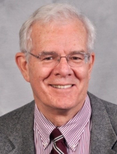
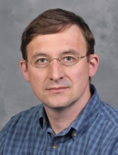
Sirotkin Lab scientific focus: My lab focuses on investigating the mechanisms of actin assembly at sites of endocytosis using spinning disk confocal microscopy and quantitative image analysis as primary tools. I am currently the director of our Department spinning disk confocal microscopy facility.

Lab scientific focus: My primary research focus is on protein dynamics in myofibrils and its relations to the myofibril assembly, maintenance and myopathies. We are interested to explore the effects of several inhibitors of the ubiquitin proteasome system (UPS), many of which are used as anticancer drugs, in order to analyze their off-target effects on muscle myofibril assembly in cardiac and skeletal muscle cells.
Lab expertise (keywords): Cardiomyocytes, Skeletal muscle, Myofibrillogenesis, FRAP, Ubiquitin Proteasome System
