Upstate researchers pioneer microscopy technique to uncover the secrets of vision loss, offering hope for new therapies
Retinitis pigmentosa (RP) is a group of inherited eye diseases that cause progressive vision loss. It’s a leading cause of visual disability and blindness in people under 60 years old. While currently there are no treatments to cure or stop the progression of vision loss, Upstate researchers have developed a new microscopy technique to better understand how the gene mutation affects processes in the eye. Peter Calvert, PhD and his team will be using a five-year, $2.8 mil grant from the National Eye Institute to observe in real time how a key protein is properly delivered and packed into new disc membranes in photoreceptors; cells that convert light into electrical signals that allow us to see. Understanding this process could help find new therapies to extend or restore vision in patients with RP and other degenerative eye diseases.
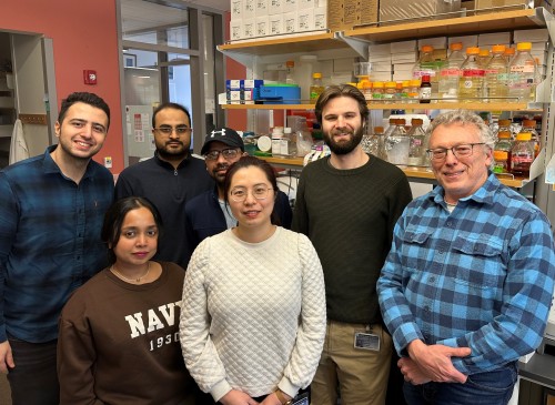
Members of the Calvert Lab, who have pioneered a microscopy technique to help study the mechanism of rhodopsin delivery. Pictured from left to right: Rasul Rakhshan, Farzana Khan, Faizan Zaidi, Himanshu Malhotra, Jiaqi Huang, Matthew Krause, Peter Calvert.
Calvert, a Professor of Ophthalmology and Visual Sciences at Upstate’s Center for Vision Research, explains that this project is focused on understanding how light-sensing structures in rod photoreceptors of the eye, called outer segments, are generated. Every day, 30 million proteins called rhodopsins are delivered to each rod outer segment in our eyes, supporting the creation of 80 new discs. While researchers know how rhodopsins are made, what happens next is one of the major unanswered questions in vision. “Before we understand the mechanism of rhodopsin delivery,” says Calvert, “it's going to be hard to find therapies for many blinding diseases.”
“Our thought is that in order to really understand how that works, we needed to watch it happen in living cells; we need to watch the process of those proteins being delivered,” explains Calvert. He and his team have developed a microscopy approach called 3D single particle tracking photoactivation-localization microscopy (3D-sptPALM). “We are going to watch rhodopsins enter the outer segment, move along the connecting cilium, and get packed into the discs.”
Calvert’s lab is one of only a handful across the country that are utilizing this technology; even fewer are using it on live tissue. By highlighting a few rhodopsins at a time with florescent markers, they can watch them travel in real time. Applying this process repeatedly in the same cell allows them to map thousands of movements. Working with an expert in mathematical modeling, they’ll create a 3D model of the molecular dynamics in a single photoreceptor. “The notion is that once we have the model, we'll have a good understanding of what features of the proteins and the cell’s structure are important for getting the molecules to be in the right place.”
Calvert highlights the personal toll RP and other blinding diseases take, and the impact a treatment would have on people facing immanent vision loss.
“I was reading recently about this family where the parents have recessive RP genes, and two of their three kids have RP. The family is traveling the world so that those kids can see everything before they're blind. Can't we get in there and fix that before those kids have to lose their sight? That's the kind of thing we're trying to do here.”
Using this technology to study the movement of rhodopsins could be just the beginning of its research applications, says Calvert.
“There are other kinds of diseases like Alzheimer's disease that also may have some sort of transport issues, so this isn't just restricted to vision and blindness. I think it has broader implications, and I’m so excited about that.”
You can read more about the research happening in the Calvert lab here-
https://www.upstate.edu/cvr/investigators/peter-d-calvert-phd/index.php